

Practice Perfect 904
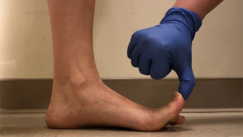
The Hubscher Maneuver: What’s the Big Deal?
The Hubscher Maneuver: What’s the Big Deal?
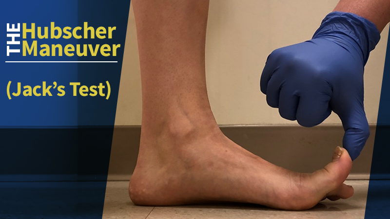
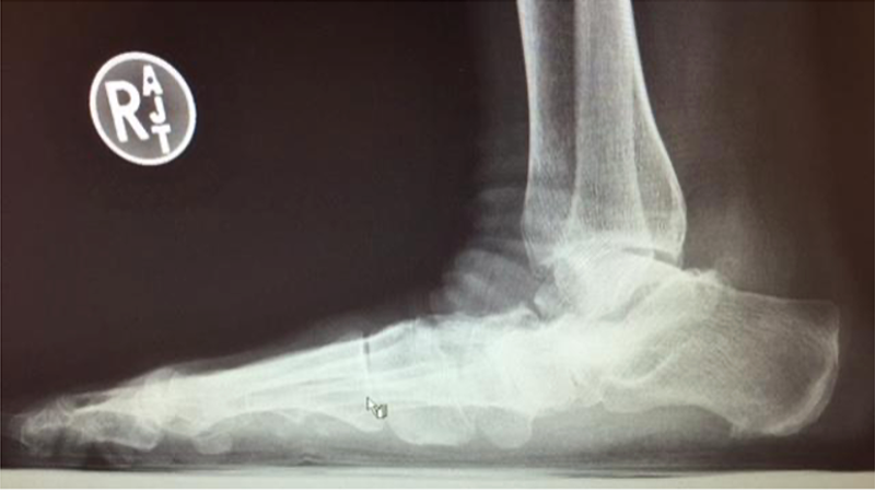
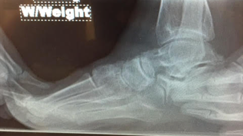
As I teach clinical podiatry to students and residents, the one physical examination maneuver they all seem to love (in the sense that they universally perform the procedure) is the Hubscher maneuver. It’s possible this is the case because it’s a simple procedure. Have the patient stand while you passively dorsiflex the great toe and see what happens. Not terribly complicated.
However, I assert most of our trainees don’t appreciate all aspects of this maneuver in terms of what it tells us about a patient’s foot function.


Recall that the Hubscher maneuver relies on the Windlass effect of the plantar fascia. Dorsiflexing the great toe winds the distal aspect of the fascia around the sesamoids and first metatarsal head, tightening the fascia, decreasing the distance from its origin to insertion, which pulls the arch up in feet that are flexible enough for this to occur.
It’s important to understand that this test has important limitations. The first is to realize that this test simulates dorsiflexion of the hallux during the propulsive phase of gait, but it only simulates it. Halstead and Redmond found no relationship between the amount of hallux dorsiflexion during the Hubscher maneuver compared with the amount seen during active gait.1 These authors advocated abandoning the Hubscher maneuver in favor of instrumented methods. Unfortunately, no one in practice has these devices, and they wouldn’t be practical for a busy office. I agree that this test is not valid for the diagnosis of hallux limitus, but the Hubscher tells us more than simply the amount of hallux dorsiflexion.
All the way back in 1953, Dr Ewen Jack discussed this maneuver, advocating for a naviculocuneiform arthrodesis.2 He described the clinical test we know today as the Hubscher maneuver (which is why it’s also called Jack’s test), but his purpose was to determine the location at which the arch fails in a pediatric flatfoot condition as well as the flexibility of the foot. If the arch was raised during the maneuver, the foot was flexible, and if it did not raise, the foot was rigid.
For those of you historically minded, I was unfortunately not able to find the etymology of the name “Hübscher”. There’s reference to an article in a now defunct Dutch journal, but it’s not written by anyone named Hübscher (presumably pronounced “Hoob-sher”). If anyone can report on who Hübscher was, please write in!
Dr Jack also pointed out another important interpretation, which I think is often missed. If the arch collapse occurs primarily at the talonavicular joint, there will not be enough leverage to recreate the arch. Don’t be confused by this. If, during your nonweightbearing examination, the arch is flexible but in relaxed stance the arch doesn’t recreate, then it’s not a rigid deformity; there’s simply more proximal pathology overpowering the plantar fascia’s ability to pull the arch up.


There’s another important aspect to consider, which is an anatomical one. In order for the Windlass effect to occur, the other ligamentous components of the medial longitudinal arch and the hindfoot must be intact. When we perform this test in a patient with intact anatomy, we see not only arch rising, but also inversion of the heel and external rotation of the tibia. Remember, where the talus goes so too goes the tibia and vice versa (because the talus is locked into the ankle mortise). If the interosseous talocalcaneal ligament was stretched, damaged, or ruptured, then the talus is no longer connected with the calcaneus, which essentially “undocks” the foot from the leg. In a case like this, even if the arch dorsiflexes, we will not see calcaneal inversion or tibial external rotation. Finding this result in a patient with a planus condition pending surgery would lead one to the conclusion that a subtalar arthrodesis is an important procedure to choose.
Clearly, there’s more to this test than simply pulling up the big toe!
Best wishes.

Jarrod Shapiro, DPM
PRESENT Practice Perfect Editor
[email protected]

-
Halstead, Jill and Anthony C. Redmond. Weight-bearing passive dorsiflexion of the hallux in standing is not related to hallux dorsiflexion during walking. J Orthop Sports Phys Ther. 2006 Aug; 36(8):550-556.
Follow this link -
Jack EA. Naviculo-cuneiform fusion in the treatment of flat foot. J Bone Joint Surg Br. 1953 Feb;35-B(1):75-82.
Follow this link
Major Sponsor


Comments
There are 0 comments for this article